Home » Uncategories » Back Muscles Anatomy / Stockfoto Labeled Anatomy Chart Of Male Back Muscles O : These muscles work together to move the scapula anteriorly and laterally during pushing, throwing, or punching motions.
Back Muscles Anatomy / Stockfoto Labeled Anatomy Chart Of Male Back Muscles O : These muscles work together to move the scapula anteriorly and laterally during pushing, throwing, or punching motions.
Back Muscles Anatomy / Stockfoto Labeled Anatomy Chart Of Male Back Muscles O : These muscles work together to move the scapula anteriorly and laterally during pushing, throwing, or punching motions.. Leaning back to straight vertical and all points in between. (2017, elsevier) should be consulted. This curve, called lordosis, helps to: The back comprises interconnecting nerves, bones, muscles, ligaments, and tendons, all of which can be a source of pain. These muscles give height and breadth to back development.
The deep muscles develop in the back called intrinsic muscles. Three types of back muscles that help the spine function are extensors, flexors and obliques. This blog post article is an overview of the muscles of the lumbar spine of the trunk. We hope this picture anatomy of back muscles diagram can help you study and research. These structures work together to support the body, enable a range of movements, and send messages from the brain to the.
Back Muscle Anatomy And Workouts To Strengthen from nitrocdn.com These muscles include the large paired muscles in the lower back, called erector spinae, which help hold up the spine, and gluteal muscles. The muscles of the back muscles make up a large part of the anatomy (structure) of the back. Muscles of the lumbar spine. Anatomy of the back muscles the latissimus dorsi muscles (also known as the lats) are the largest muscles of the back. The superficial back muscles are situated underneath the skin and superficial fascia. The multifidus, a long muscle that travels nearly the entire length of the back.it helps to stabilize and rotate the lower back, and additionally takes some. The back comprises interconnecting nerves, bones, muscles, ligaments, and tendons, all of which can be a source of pain. The superior part of the appendicular skeleton that includes clavicle, scapula, and humerus, is attached to the axial skeleton that consists of skull.
Anatomy chart courtesy of fcit the latissimus dorsi muscles (also known as the lats) are the largest muscles of the back.
Your lower back (lumbar spine) is the anatomic region between your lowest rib and the upper part of the buttock. They provide movements of the spine , stability to the trunk, as well as the coordination between the movements of the limbs and trunk. The superficial back muscles are situated underneath the skin and superficial fascia. These layers of back muscles help to mobilize and stabilize your trunk during your day to day activities. By far the most common cause of back pain is muscle strain. Superficial back muscles, intermediate back muscles and intrinsic back muscles.the intrinsic muscles are named as such because their embryological development begins in the back, oppose to the superficial and intermediate back muscles which develop elsewhere and are therefore classed as extrinsic muscles. The back comprises interconnecting nerves, bones, muscles, ligaments, and tendons, all of which can be a source of pain. These muscles determine body posture and also regulate the three basic movements of the trunk: All about the back muscles the back anatomy includes the latissimus dorsi, trapezius, erector spinae, rhomboid, and the teres major. Superficial muscles of the back are located directly deep towards the skin along with superficial fascia.they are occasionally called the appendicular group as these muscles are mainly associated with activities of the appendicular skeleton. We hope this picture anatomy of back muscles diagram can help you study and research. The intrinsic back muscles are found deeper to the extrinsic muscles, separated from them by the thoracolumbar fascia. Bodybuilder showing his back and biceps muscles, personal fitness trainer.
They provide movements of the spine , stability to the trunk, as well as the coordination between the movements of the limbs and trunk. They start at the top of the neck and go down to the tailbone. The superior part of the appendicular skeleton that includes clavicle, scapula, and humerus, is attached to the axial skeleton that consists of skull. These muscles determine body posture and also regulate the three basic movements of the trunk: The muscles of the back can be arranged into 3 categories based on their location:
Back Muscles Attachments Nerve Supply Action Anatomy Info from anatomyinfo.com The extrinsic back muscles are located in the back, but act to produce movements of the shoulder and assist respiration. Back pain is common and might be caused by a problem with a muscle. Back pain is one of the most common kinds of pain for adults, and muscle strains are the most common type of back pain. Balance the weight of your head on top of your spine evenly distribute weights from your upper body into the lower extremities These layers of back muscles help to mobilize and stabilize your trunk during your day to day activities. Muscles of the lumbar spine. Anatomy of back muscles your back consists of three distinct layers of muscles, namely the superficial layer, the intermediate layer, and the deep layer. The human spine is composed of 4 sections of vertebrae.
The human spine is composed of 4 sections of vertebrae.
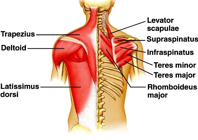
These structures work together to support the body, enable a range of movements, and send messages from the brain to the. In the upper back region, the trapezius, rhomboid major, and levator scapulae muscles anchor the scapula and clavicle to the spines of several vertebrae and the occipital bone of the skull. Back pain is the second most common type of pain in adults (the most common being headaches). They start at the top of the neck and go down to the tailbone. What are the lower back muscles and their anatomy? These muscles give height and breadth to back development. The human spine is composed of 4 sections of vertebrae. The back muscles are anatomically layered into superficial (extrinsic) and deep (intrinsic) muscles. These layers of back muscles help to mobilize and stabilize your trunk during your day to day activities. Anatomy chart courtesy of fcit the latissimus dorsi muscles (also known as the lats) are the largest muscles of the back. The superficial back muscles are situated underneath the skin and superficial fascia. We think this is the most useful anatomy picture that you need. All these muscles are therefore associated with movements of the upper limb.
Muscles that act on the back. Memorize all the muscle facts with the help of muscle cheat sheets. Tutorials on the anatomy and actions of the back muscles, using interactive animations, diagrams, and illustrations. In the upper back region, the trapezius, rhomboid major, and levator scapulae muscles anchor the scapula and clavicle to the spines of several vertebrae and the occipital bone of the skull. The muscles of the back muscles make up a large part of the anatomy (structure) of the back.
The Ultimate Guide To Back Spasms from www.spineuniverse.com Anatomy of the back muscles the latissimus dorsi muscles (also known as the lats) are the largest muscles of the back. The muscles of the lower back help stabilize, rotate, flex, and extend the spinal column, which is a bony tower of 24 vertebrae that gives the body structure and houses the spinal cord.the spinal. The back muscles are divided into two large groups: These muscles include the large paired muscles in the lower back, called erector spinae, which help hold up the spine, and gluteal muscles. Deep muscles of the lower back include: Muscle or ligament strains can occur from repeated use of the muscles, or from improperly or awkwardly lifting heavy objects. These sections are cervical (neck), thoracic (upper and middle back), lumbar (lower back), and sacrum (tailbone). The superior part of the appendicular skeleton that includes clavicle, scapula, and humerus, is attached to the axial skeleton that consists of skull.
The superficial back muscles are situated underneath the skin and superficial fascia.
What are the lower back muscles and their anatomy? We think this is the most useful anatomy picture that you need. Back pain is the second most common type of pain in adults (the most common being headaches). They start at the top of the neck and go down to the tailbone. These muscles give height and breadth to back development. The muscles of the back muscles make up a large part of the anatomy (structure) of the back. These sections are cervical (neck), thoracic (upper and middle back), lumbar (lower back), and sacrum (tailbone). Back muscles, functions and exercises: Muscle or ligament strains can occur from repeated use of the muscles, or from improperly or awkwardly lifting heavy objects. Anatomy chart courtesy of fcit the latissimus dorsi muscles (also known as the lats) are the largest muscles of the back. The back muscles are divided into two large groups: These muscles determine body posture and also regulate the three basic movements of the trunk: Tutorials on the anatomy and actions of the back muscles, using interactive animations, diagrams, and illustrations.
0 Response to "Back Muscles Anatomy / Stockfoto Labeled Anatomy Chart Of Male Back Muscles O : These muscles work together to move the scapula anteriorly and laterally during pushing, throwing, or punching motions."


0 Response to "Back Muscles Anatomy / Stockfoto Labeled Anatomy Chart Of Male Back Muscles O : These muscles work together to move the scapula anteriorly and laterally during pushing, throwing, or punching motions."
Post a Comment